Secretion assay


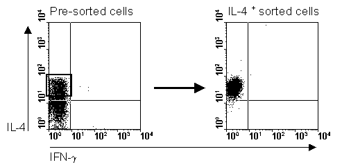
Manz et al. (1995) developed  the secretion assay, which makes it possible to detect secreted cytokines in an elegant way. Cells are coated with an antibody-antibody complex that coats the cell and is able to catch (by the cell) secreted molecules. The secreted cytokine is detected by a second antibody labelled with a fluorochrome. Bosterhus et al. (1999) took it a step further and adjusted the protocol that makes it is possible to sort these cells by a magnetic based separation system or in a flow cytometer with sort option. This way it is possible to continue your culture based on the secretion of the molecule and the detection of the fluorochrome labelled antibody. The technique is comparable with a cell surface based ELISA. The big advantage of this system is that you still have still living cells after the whole procedure. 

Fig.1. CD4+ T cells stimulated for 3 hours with PMA/ionomycin, labeled with the secretion assay, 15 min. secretion time (left part of figure) and sorted on a FACSVantage (right part of figure) Published in: H.H.Smits et al. Eur.J.Imm. Vol 31, 1055 - 1065 (2001)
 

Presentation on 'Isolation and detection of specific cytokine producing cells by MACS Cytokine Secretion Assay' Blijdorp, R'dam, April 10th 2001.

Although we did our best we can not be held responsible for any mistakes or typing errors.
 Suggestions, literature and protocols are welcome at icstaining@schuitemaker.info. Copyright 2000 - 2005.
Page was last updated: 05-03-2005