 BrdU
BrdU
![]()
![]()
|
|
|
|
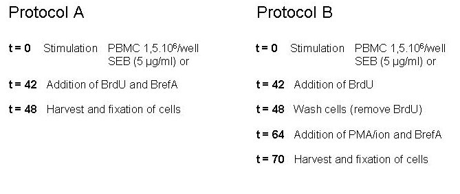
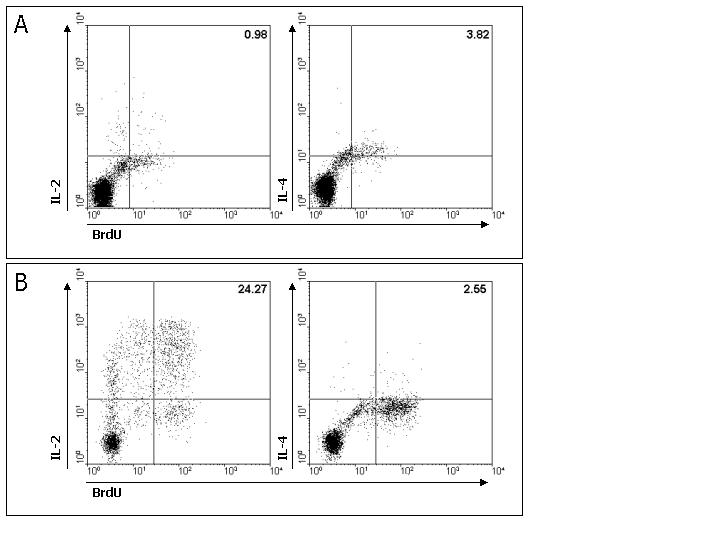
DNA synthesis by (sub)populations of cells can be tracked by making use of radioactive form of thymidine, but it can also be done by the use of its analogue 5-bromo-2-deoxyuridine (BrdU). In the newly synthesized DNA thymidine is (partly) replaced by BrdU, which is in high concentration added to the culture. After fixation and permeabilization of the cells the BrdU can be shown by making use of a fluorochrome labelled antibody against the BrdU. Using DNAse during the staining period the BrdU becomes more available to the antibody without influencing the fluorescence or the structure of the cell. One company is bringing a kit that makes it possible to have a balanced presence of antibody and DNAse in the sample. Several publications are available comparing the method to thymidine or using BrdU together with surface markers. Both in polyclonal and antigen specific stimulation. We tried to enhance the protocol for the detection of cytokines
together with the BrdU by adding an
extra step. In the figure below a comparison is made between the standard
protocol and ours. After the 'regular' 48 hrs we wash away the BrdU, that was
added after 42 hours, and incubated the cells for an other 16 hrs (over
night) and stimulated them with PMA/ionomycin for 6 hrs (total time is 70 hours
now). During the last 4 hrs
Brefeldin A (BFA) was present (table.1.). The idea is that the initial stimulation shows the
reactive antigen cells in the population. These will be BrdU positive. And the
PMA/ionomycin will show the cytokine profile of these cells. The number of cytokine producing cells is clearly enhanced. And
also the total number of BrdU positive cells is larger (fig.1.).
|
|
Although we did our best we can
not be held responsible for any mistakes or typing errors. |